Keywords
Glial Fibrillary Acidic Protein, Ubiquitin C-Terminal Hydrolase L1, Traumatic Brain Injury, Reference Values, Paediatrics, Biomarkers, Immunoassay
Glial Fibrillary Acidic Protein, Ubiquitin C-Terminal Hydrolase L1, Traumatic Brain Injury, Reference Values, Paediatrics, Biomarkers, Immunoassay
Over one million people attend UK emergency departments annually with mild traumatic brain injury (mTBI).1 Approximately 33–50% of these are children and adolescents, many of whom will receive computerised tomography (CT) scans to rule out intracranial lesions (ICLs). Only a small proportion of children who undergo a CT scan for mTBI will show ICLs, however, these require an accurate diagnosis as ICLs are associated with an increased risk of secondary insults and poorer outcomes.2 Despite the clear diagnostic benefits of CT scans, their use in children poses a dilemma due to high costs and radiation risks, with cancer risk now thought to be greater than previously estimated.3 This is especially challenging for intermediate-risk mTBI cases, where many CT scans ultimately show normal findings.
Blood biomarkers have shown promise as risk stratification tools to identify patients at very low risk of ICLs who can safely avoid CT imaging.4 Glial fibrillary acidic protein (GFAP) and ubiquitin C-terminal hydrolase L1 (UCH-L1) are brain-specific proteins released into circulation following astroglial and neuronal injury, respectively.5,6 These biomarkers have been developed into commercial clinical assays by Abbott (i-STAT TBI Plasma test) and bioMérieux (VIDAS® TBI (GFAP, UCH-L1)). Diagnostic accuracy studies in adults, along with characterisation of biomarker concentrations in healthy populations, have demonstrated their utility in the evaluation of mTBI, and both assays have defined cut-off values established for use in adults. The bioMérieux VIDAS® TBI (GFAP, UCH-L1) assay is CE-marked in the European Union and has received U.S. FDA 510(k) clearance for use in adults.7
Studies investigating GFAP and UCH-L1 in children for ruling out CT imaging are more limited but show promising diagnostic accuracy.8–12 There is also limited knowledge of GFAP and UCH-L1 paediatric reference intervals (RIs) in healthy populations. Accurate RIs are essential for developing clinical decision thresholds, which are needed before these assays can be validated and implemented for children. Paediatric RIs have been reported in one French substudy for the Abbott assay, but not for the bioMérieux platform, and they have not yet been confirmed in an international population.12 Given the substantial brain development and maturation occurring in early childhood, and existing evidence of age-related variation in GFAP and UCH-L1 concentrations in adults, age-specific thresholds are likely to be required for paediatric populations. Indeed, several previous investigations have identified statistically significant age-related partitions for GFAP across different assays, further supporting this need.12–14
This study aimed to establish paediatric RIs for GFAP and UCH-L1 and to determine the need for age- or sex-specific interpretation. These findings will support ongoing work to validate this assay for paediatric clinical use.
Patients and the public were involved in the design of this research. Thirty parents, children and young people were consulted about the acceptability of blood-based biomarkers to reduce CT scanning following head injury; there was unanimous support for a reliable blood test to help avoid unnecessary radiation exposure, and 87% favoured the use of stored blood samples from healthy children to establish reference values to minimise additional painful procedures. A lay summary of findings will be co-developed with the PERUKI Young Persons Advisory Group to support dissemination to participants and wider communities.
This study was conducted in accordance with Clinical and Laboratory Standards Institute (CLSI) guideline EP28-A3c for the establishment of reference intervals.17 Serum samples were taken from a cohort of healthy children, collected as part of the Rapid Diagnostics Antibody Testing and Host Response in Children with COVID-19 (RAPID-19) study, previously registered at ClinicalTrials.gov NCT04347408.15,16 This trial was registered on 15th April 2020, participant enrolment began on 16 April 2020 and was completed on 3rd July 2020. Blood samples and basic demographic data (age, sex, site) were collected from children aged 2–16 years with no symptoms of illness or recent hospital attendances for any reason at 5 UK sites.
Sample selection was guided by CLSI EP28-A3c recommendations, which advise a minimum of 120 individuals per partition. Based on existing paediatric GFAP literature, three age categories were prespecified (2–<4, 4–<11, and 11–<16 years). Samples were selected to achieve n ≥ 120 in each age group where possible, or otherwise to include the maximum number of available specimens. Sex and recruitment site were balanced across age groups.
Blood was collected by venepuncture and processed as serum according to local site standard operating procedures, full details of sample collection have previously been described.16 After collection, serum aliquots were taken and stored at −80 °C until required for sample analysis. Serum samples used in this study had a maximum of two freeze-thaw cycles.
Ethical approval for the original RAPID-19 study was obtained from the London-Chelsea Research Ethics Committee (REC reference: 20/HRA/1731) and the Belfast Health & Social Care Trust Research Governance (reference: 19147TW-SW). Ethical approval to use the stored samples for the present study was obtained from the Queen’s University Belfast Faculty of Medicine, Health and Life Sciences Research Ethics Committee (reference: MHLS 24_136). Written informed consent was obtained from all participants’ parents or legal guardians, and age-appropriate assent was obtained from the children where possible. Participants were free to decline or withdraw consent at any time without providing a reason and without any resulting detriment.
Samples were thawed and analysed immediately using the BioMérieux VIDAS® TBI (GFAP, UCH-L1) assay on VIDAS® KUBE™ Immunoassay Analyzer (bioMérieux, France), according to the manufacturer’s instructions. The assay employs an automated immunoassay. Results were automatically calculated and expressed in VIDAS® test values using the system’s built-in calibration. All samples were randomised and analysed blind to participant demographics and clinical data.
Assay performance was monitored using manufacturer-provided quality control materials, as per the instrument’s predefined acceptance criteria, before sample testing proceeded. Between-run CVs, determined from repeated analysis of a single serum sample over multiple runs, were 2.7% for GFAP and 4.7% for UCH-L1, respectively. No sample was above the upper range limit, samples below-LoD were assigned as the relative LoD (10 pg/ml for GFAP, 80 pg/ml for UCH-L1.
Establishment of reference intervals for GFAP and UCH-L1 took place according to the approved guideline set out by the CLSI EP28-A3c guidelines.17 Removal of extreme outliers took place by inspection of scatter and distribution plots of concentration against age. The Harris and Boyd method was used to detect any statistically significant age or sex partitions, before data in each partition was transformed using the Box-Cox method. Data normality of each identified partition was assessed using Q-Q plots and the Shapiro-Wilks test and outliers were removed from the partitioned data using the Tukey method for normal data and the adjusted Tukey test twice for skewed partitions. Lower and upper RIs and equivalent 90% confidence limits were calculated using the non-parametric percentile method when n ≥ 120 and using the Horn and Pesce robust method in any partition in which n < 120.
As GFAP varied continuously with age, a regression-based approach was also used, as well as discrete age partitioning. Continuous age-based reference intervals were determined using weighted polynomial regression. 90% confidence intervals for the centiles were obtained using 2,000-iteration bootstrap resampling. Analyses were performed using Microsoft Excel and MedCalc® software version 22.026.
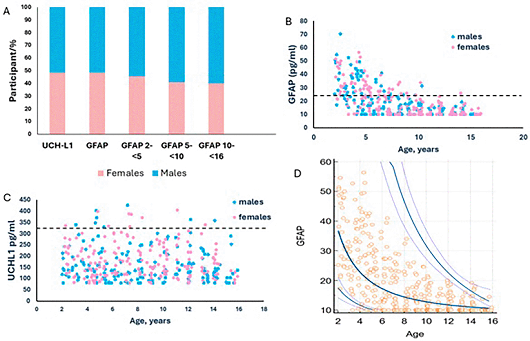
This study included 370 children aged 2–16 years, recruited across five UK sites. 190/370 (51.4%) were males and 180/370 (48.6%) females, with a median age of 9.8 years (IQR 8.2–11.96 years). Samples were from five UK sites: Site 1, Belfast n = 77 (20.8%), Site 2, Glasgow n = 64 (17.3%), Site 3, London n = 100 (27%), Site 4, Manchester n = 72 (19.5%), and Site 5, Cardiff n = 57 (15.4%). GFAP and UCH-L1 concentrations were measured in all serum samples, except in 11 cases where sample volume was insufficient for UCH-L1 analysis. Demographic characteristics and the distribution of participants by sex are summarised in Table 1 and Figure 1A.
. Demographic characteristics of the study population. Age groups, sex distribution, and recruitment numbers by study site for participants with available GFAP and UCH-L1 measurements.
. Distribution Patterns and Age-Dependent Reference Intervals for Pediatric GFAP and UCH-L1 Biomarkers.
(A) Distribution of study participants by sex for each biomarker. For GFAP, sex distribution by age group (2–<5, 5–<10, and 10–<16 years) relevant to reference-interval determination is also shown.
(B–C) Scatterplots of GFAP (pg/mL) (B) and UCH-L1 (pg/mL) (C) concentrations by age (years). Data points are coloured by sex. Horizontal dashed lines indicate bioMérieux assay cutoffs for traumatic brain injury indication in adults (GFAP: 22 pg/mL; UCH-L1: 327 pg/mL).
(D) Continuous prediction reference intervals for the age-related biomarker GFAP. The curves represent the calculated mean concentration and the 2.5th and 97.5th centiles.
Examination of boxplots and scatter graphs identified outliers for both GFAP (n = 1) and UCH-L1 (n = 3) (Supplemental Figure 1). Visual inspection of the scatterplots revealed a continuous age-related change in GFAP concentrations (Figure 1B). The need for age partitioning of GFAP was further confirmed using the Harris and Boyd method. Although GFAP levels increased progressively and continuously with age, for clinical interpretability, discrete age groups were selected based on visual assessment of the data and verified by Harris and Boyd: 2–<5 years (n = 95), 5–<10 years (n = 134), and 10–<16 years (n = 140). No age-related change in UCH-L1 concentrations was observed (Figure 1C). Assessment of scatterplots and application of the Harris and Boyd approach indicated that sex partitioning was not required for either biomarker, and this finding was consistent across all GFAP age groups.
Reference intervals were determined for each GFAP age group and for UCH-L1. Following Box–Cox transformation, Gaussian distributions were achieved for UCH-L1 and for the GFAP 2–<5-year group, whereas the remaining GFAP age groups retained skewed distributions. Additional outliers were removed prior to final reference-interval estimation (GFAP 2–<5 years: n = 1; GFAP 10–<16 years: n = 4). Upper reference intervals were calculated as the 97.5th percentiles, with corresponding 90% confidence intervals, and are presented in Table 2.
. Normative upper reference interval for GFAP and UCH-L1. Upper (97.5th) reference limits are shown with their corresponding age range and sample size.
Reference intervals were defined for the three GFAP age groups and UCH-L1. After Box Cox transformation, Gaussian distribution was obtained for the UCH-L1 and GFAP 2- < 5 years datasets, but the other two datasets remained skewed. Prior to final reference interval determination, further outliers were identified and removed (GFAP, 2- < 5 years, n = 1; GFAP, 10- < 16 years, n = 4). Reference intervals for all markers and partitions were calculated as 2.5th and 97.5th percentiles and are displayed alongside corresponding 90% confidence intervals in Table 2.
Applying the BioMérieux adult TBI cutoffs after final outlier removal would have yielded 17/356 (4.8%) false-positive results for UCH-L1. For GFAP, false-positive rates varied markedly by age group: 2–<5 years: 68/94 (72.3%), 5–<10 years: 38/134 (28.4%), and 10–<16 years: 0/136 (0%).
To further characterize the continuous age-related variation in GFAP concentrations, we derived age-dependent continuous reference intervals using weighted polynomial regression (Figure 1D). The upper (97.5th) centile demonstrated a pronounced decline from early childhood into adolescence. Additionally, the spread of GFAP concentrations decreased with increasing age, with the widest 90% confidence intervals observed at younger ages and progressive narrowing across older ages. These age-specific estimates and their confidence intervals are provided in Table 3.
. Continuous reference interval estimates for GFAP. Lower and upper limits, (5th and 95th percentiles) with corresponding 90% CI values for the age-associated markers GFAP (pg/ml) are shown. Determined using weighted polynomial regression.
In this study, we established paediatric reference intervals for GFAP and UCH-L1 measured using the bioMérieux VIDAS® TBI assay in a large, well-characterised cohort of healthy UK children aged 2–16 years. These data fill an important evidence gap, as paediatric reference intervals have not previously been defined for this platform and remain limited for other assays. Given ongoing efforts to evaluate blood biomarkers as tools to reduce unnecessary CT imaging in children with mild traumatic brain injury, accurate age-appropriate reference values are essential for future diagnostic validation and implementation.
GFAP demonstrated a clear and continuous age-related pattern, with concentrations highest in early childhood and gradually declining into adolescence. This trajectory is consistent with previous studies describing developmental changes in GFAP expression and turnover, and with earlier paediatric work using the Abbott platform, which also reported higher GFAP concentrations in younger children.12–14,18
Although GFAP varied continuously with age, we identified three discrete age partitions for clinical usability. Both the partitioned reference intervals and the regression-derived continuous intervals highlight the marked elevation of GFAP in early childhood compared with older children. Importantly, applying the adult BioMérieux TBI cut-off (22 pg/mL) resulted in high rates of false positives in younger age groups, over 70% in children aged 2–<5 years and nearly 30% in those aged 5–<10 years.
In contrast to GFAP, UCH-L1 showed no meaningful variation with age or sex in this cohort, consistent with reports from paediatric studies using other assays.12,14 Application of the adult BioMérieux cut-off for UCH-L1 identified a small proportion of children as potential false positives (4.8%), suggesting that while adult thresholds may be closer to suitable for paediatric use than for GFAP, further optimisation is still warranted, particularly given the low prevalence of CT-detectable intracranial lesions and the corresponding need for very high specificity in paediatric mTBI screening.
Our findings therefore align with the limited existing paediatric literature, including the French substudy of the Abbott assay, which similarly reported age-related GFAP variation and relatively stable UCH-L1 concentrations across childhood.12 By providing reference values derived using a different analytical platform and from a geographically distinct population, our results confirm that age-related variation persists across assays and must be accounted for when interpreting GFAP levels in children.
Strengths of this study include adherence to CLSI EP28-A3c guidelines, rigorous outlier assessment, and the use of both partition-based and regression-based approaches to characterise age effects. The multi-site UK recruitment enhances generalisability, and sample sizes exceeded CLSI recommended thresholds in all partitions, apart from GFAP 2–<5 years. A key strength of this study is the use of a cohort specifically recruited as healthy children, rather than relying on convenience samples obtained from children undergoing blood tests for unrelated outpatient reasons, as is common in reference-interval research.12 This reduces the likelihood that biomarker concentrations were influenced by underlying medical conditions or clinical indications for testing, thereby increasing confidence that the observed values truly reflect healthy physiological ranges.
Several limitations should be acknowledged. While this study establishes paediatric reference intervals, diagnostic decision limits for mTBI cannot be inferred from these data and require separate clinical validation in injured populations. For GFAP, the 2–<5 year partition contained fewer than the n = 120 samples than recommended by the CLSI EP28-A3c guidelines.17 Although the Horn and Pesce robust method was used and the available sample size (n = 94) was only slightly below the criterion, this remains a methodological limitation. In addition, children <2 years of age were not included, limiting generalisability to this age group. Previous work, including aged 6 months–<2 years, has shown that this group require different GFAP cut-off values, underscoring the need for caution when extrapolating our findings to younger children.12
The wide 90% confidence intervals around the upper centiles of the continuous age-dependent GFAP reference intervals likely reflect the substantial biological variability seen in younger children. Although a limited sample size in this age range may contribute to uncertainty, our findings also highlight that establishing a single fixed cut-off value for GFAP in early childhood may be inherently unreliable. Applying uniform thresholds in children under approximately 6–8 years may risk misclassification because of rapidly changing age-related physiology.
This study provides the first paediatric reference intervals for GFAP and UCH-L1 using the bioMérieux VIDAS® TBI assay, demonstrating pronounced age dependence for GFAP and relative stability of UCH-L1 across childhood. These findings highlight the limitations of applying adult thresholds to paediatric populations and establish a necessary foundation for biomarker-guided assessment of suspected mTBI in children. The wider variability observed in younger age groups underscores the need for age-specific interpretation, and future diagnostic accuracy studies should ensure adequate representation of young children to refine and validate appropriate decision limits for clinical use.
CM and TW were involved in the conception, design and obtainment of funding of this study. TW was the PI of the original RAPID-19 study. NS and OE ran the sample analysis which was verified by CM. CM and DC undertook and verified the data and statsicial analysis. DC wrote the first draft of the report with input from CM. All authors reviewed and approved the final manuscript and had full access to all the data in the study and had final responsibility for the decision to submit for publication.
Artificial intelligence (ChatGPT by OpenAI) was used to assist with improving the clarity, grammar, and readability of the manuscript. All AI-generated content integrity. No AI tools were used for data analysis or interpretation. The authors take full responsibility for the content of the published article.
De-identified participant data for this study is available on the Queen’s University Belfast data repository. This data is licenced under a CC BY licence. Please cite Mills, C, Waterfield, T. (2026). Dataset for “Age-specific Reference Intervals for Serum GFAP and UCH-L1 in Children and Adolescents”. Queen’s University Belfast. DOI: 10.17034/bf63a196-bead-4c67-93c5-8470d3420d63.
Provide sufficient details of any financial or non-financial competing interests to enable users to assess whether your comments might lead a reasonable person to question your impartiality. Consider the following examples, but note that this is not an exhaustive list:
Sign up for content alerts and receive a weekly or monthly email with all newly published articles
Register with NIHR Open Research
Already registered? Sign in
If you are a previous or current NIHR award holder, sign up for information about developments, publishing and publications from NIHR Open Research.
We'll keep you updated on any major new updates to NIHR Open Research
The email address should be the one you originally registered with F1000.
You registered with F1000 via Google, so we cannot reset your password.
To sign in, please click here.
If you still need help with your Google account password, please click here.
You registered with F1000 via Facebook, so we cannot reset your password.
To sign in, please click here.
If you still need help with your Facebook account password, please click here.
If your email address is registered with us, we will email you instructions to reset your password.
If you think you should have received this email but it has not arrived, please check your spam filters and/or contact for further assistance.
Comments on this article Comments (0)