Keywords
Myotonic dystrophy type 1, neuromuscular, dysphagia, swallowing, quantitative muscle ultrasound (QMUS), videofluoroscopic swallowing study (VFSS)
Myotonic dystrophy type 1, neuromuscular, dysphagia, swallowing, quantitative muscle ultrasound (QMUS), videofluoroscopic swallowing study (VFSS)
Neuromuscular diseases are a heterogeneous group of conditions affecting the peripheral nervous system, which connects the brain and spinal cord to the rest of the body. The main features are muscle weakness and wasting due to damage to the nerves, muscles, or neuromuscular junction.1 Swallowing difficulties (dysphagia) are particularly prevalent in certain neuromuscular disease subtypes, typically resulting from progressive damage to nerves or muscles in the bulbar system. However, growing evidence suggests that central nervous system involvement may also play a role.2 Myotonic dystrophy type 1 (DM1) is one such subtype — a progressive, autosomal dominant disorder that affects multiple body systems and can ultimately lead to premature death. Individuals with DM1 develop myotonia (delayed muscle relaxation)3 and experience impairments in both the central and peripheral nervous systems,4,5 all of which can disrupt swallowing. In DM1, swallowing problems occur within the context of cardiorespiratory decline, cognitive impairment, and physical deterioration.6 This multisystem involvement makes individuals with DM1 particularly vulnerable to the serious health consequences of dysphagia, including aspiration pneumonia — a potentially fatal complication caused by food or liquid entering the airway. Alsaggaf et al. demonstrated this in a retrospective analysis of 1,021 UK-based patients and 15,104 control records, identifying a 15.8-fold increased risk of death from aspiration pneumonia in people with DM1 compared to sex- and age-matched controls.7 Despite this significant mortality risk, the pathophysiological mechanisms underlying dysphagia in DM1 remain poorly understood.
Identifying targeted treatments requires a deeper understanding of the pathophysiological mechanisms underlying dysphagia in DM1. The current assumption that dysphagia in DM1 results from muscle wasting has been neither confirmed nor adequately tested. Existing research has emphasised symptomatic and functional assessment, at the expense of underlying mechanisms.8 This knowledge gap prevents a comprehensive understanding of dysphagia in DM1 and its broader relationships with function and patient experience. Implementing tools that evaluate swallowing pathophysiology could facilitate phenotype-specific dysphagia treatments, which is crucial in this era of disease-modifying therapies.9 This study investigates the multidimensional profile of dysphagia in DM1 through examination of morphometric changes in swallowing-related muscles and their relationships with biomechanical function, swallowing performance, patient experience, and other disease-affected systems.
The study represents the first comprehensive investigation of the pathophysiological underpinnings of dysphagia in DM1, with the aim of producing evidence-based recommendations to guide targeted assessment and treatment.
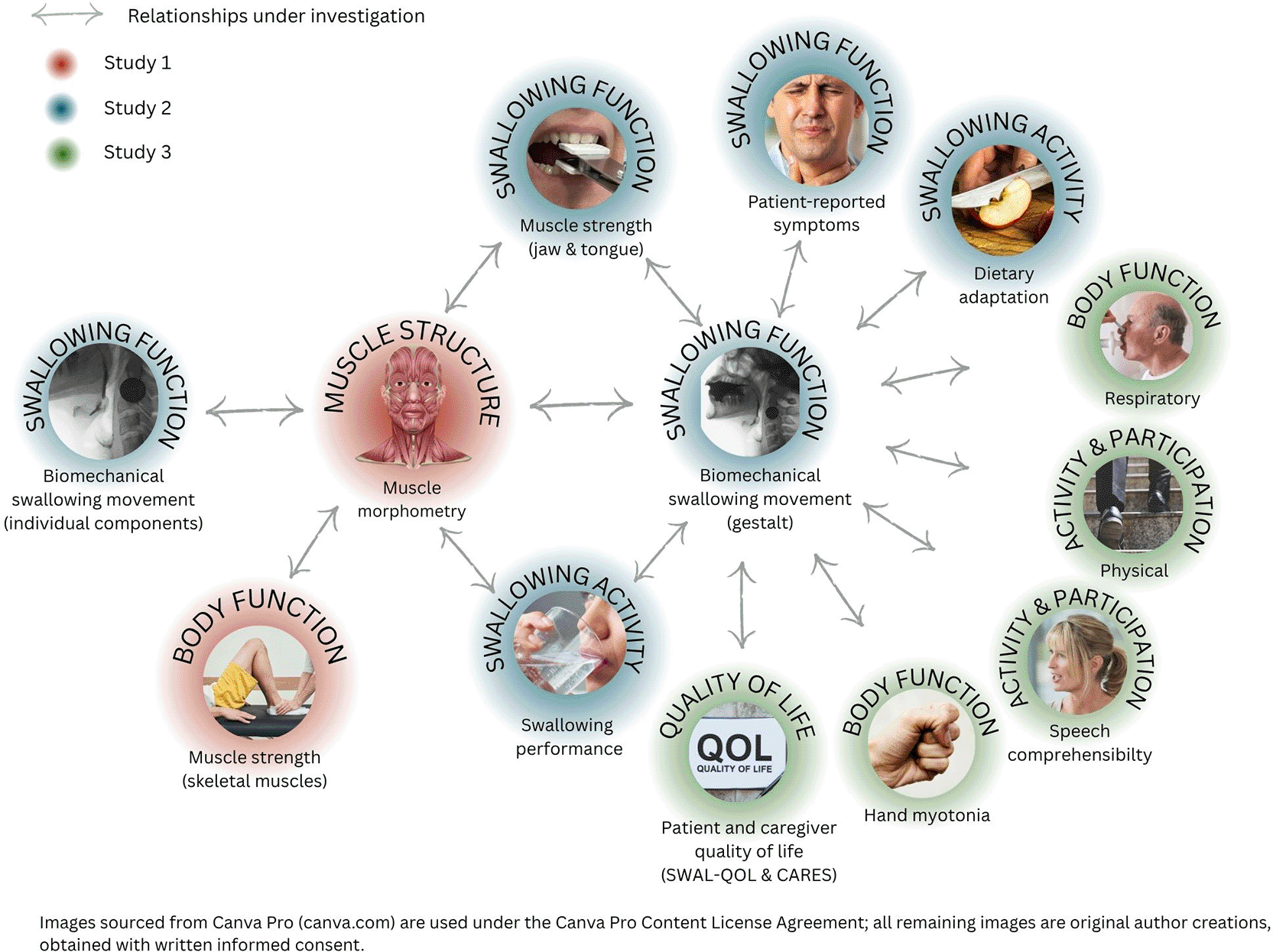
This study aims to characterise the multidimensional profile of dysphagia in people with myotonic dystrophy type 1 (pwDM1) through three integrated sub-studies:
• Sub-study one will explore the pathophysiological mechanisms associated with dysphagia in DM1 by comparing swallowing-related muscle morphometry between pwDM1 and healthy controls.
• Sub-study two will examine how underlying muscle morphometry relates to biomechanical swallowing function, swallowing-related strength and performance, and patient-reported symptoms, and explore relationships between these swallowing parameters themselves.
• Sub-study three will investigate whether biomechanical swallowing function is associated with quality of life, physical activity, and other systemic disease manifestations of DM1.
An overview of the relationships to be explored across each sub-study is presented in Figure 1.1.
Images sourced from Canva Pro (canva.com) are used under the Canva Pro Content License Agreement; all remaining images are original author creations, obtained with written informed consent.
To compare the morphometry of key swallowing-related muscles — specifically the size (thickness) and structure (echogenicity) — between pwDM1 and age- and sex-matched healthy controls.
To:
• describe the influence of sex, age, and body mass index on muscle size and structure in both pwDM1 and healthy controls;
• explore the differences among swallowing-related muscles;
• evaluate the inter- and intra-rater reliability of quantitative muscle ultrasound (QMUS) data acquisition and extraction; and
• examine the relationship between magnetic resonance imaging (MRI)-quantified fat fraction and QMUS echogenicity.
To examine the relationship between swallowing-related muscle morphometry and biomechanical swallowing function.
This study has been designed to comprehensively evaluate dysphagia across multiple domains of the International Classification of Functioning, Disability and Health (ICF) framework, encompassing body structure and function, activity, participation, and quality of life.10,11 The cross-sectional design enables efficient recruitment of an adequately powered sample within the 36-month funding period while allowing examination of relationships among multiple disease-related variables and minimising participant burden.
The protocol comprises three integrated sub-studies, each with specific aims and objectives detailed above and summarised in Figure 1.2.
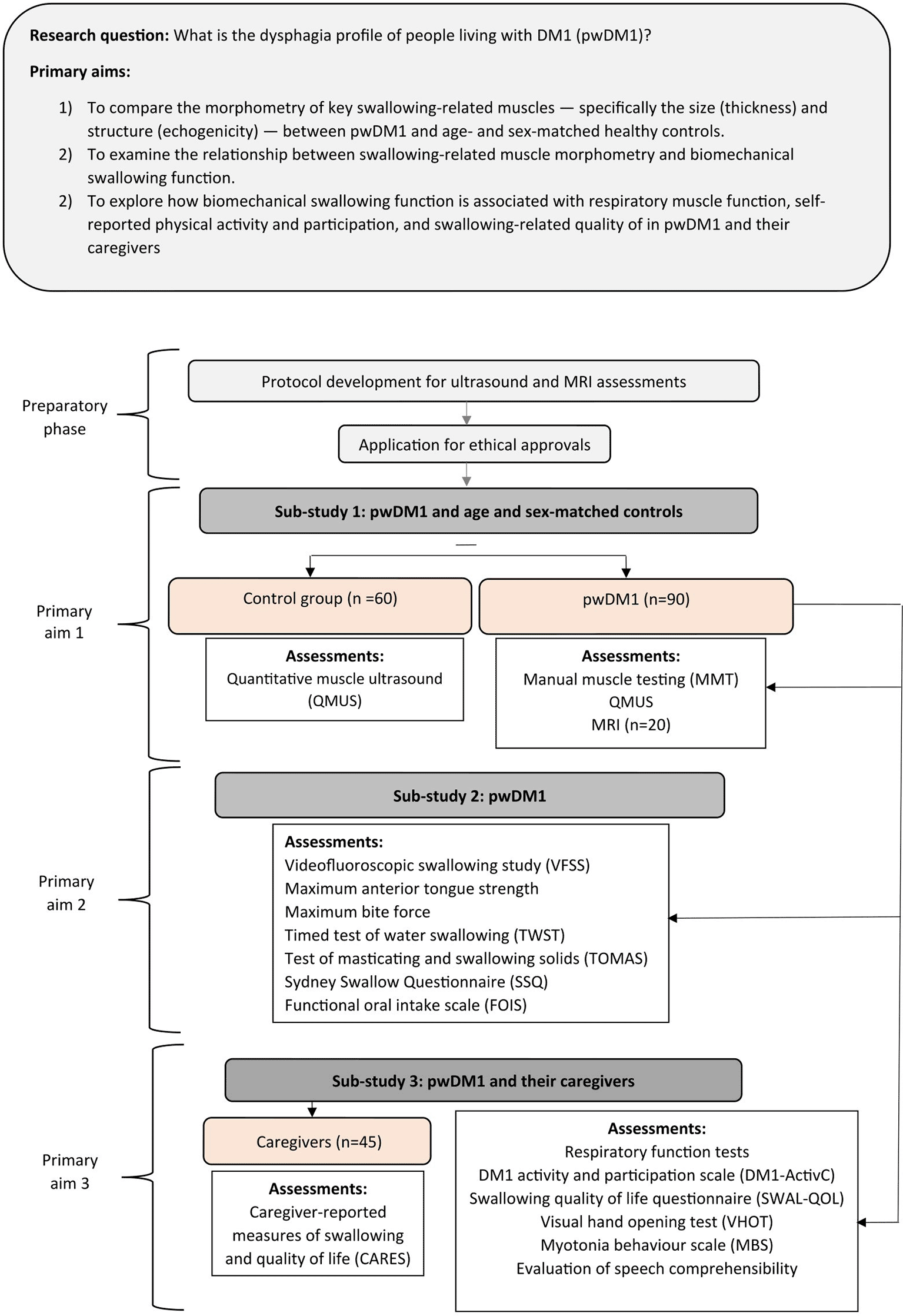
Adults with DM1 (with and without self-reported swallowing difficulties) and their caregivers will be invited to participate. To ensure representation across the full range of neuromuscular disease severity, participants will be recruited into one of three groups: mild, moderate, or severe. Group allocation will be determined by neuromuscular disability level, measured by the Muscle Impairment Rating Scale (MIRS).12 Pre-screening questions regarding muscle function will be used to facilitate balanced recruitment across all severity groups and will be integrated into the participant screening form. Age- and sex-matched healthy controls will be invited to participate to provide reference values where normative data are not currently available in the published literature. Caregivers will be invited to participate in sub-study three, in accordance with its specific aims.
Participants with DM1
Inclusion
• Established diagnosis of DM1 evidenced by genetic report;
• ≥18 years of age;
• Neuromuscular symptom onset between 14 years and 40 years; and
• Capable of consuming a minimum of five boluses of food or liquid in a single sitting.
Exclusion
• Any unrelated condition or treatment that may influence swallowing muscle composition and/or function (for example, a history of stroke or throat cancer);
• Current pregnancy;
• Facial hair under the chin and jaw line >3 mm in length (may interfere with ultrasound probe contact);
• Enrolled in disease-modifying drug trials; and
• Insufficient English language proficiency to follow test instructions.
Healthy controls
Inclusion
Exclusion
• Any condition or treatment that may influence swallowing muscle composition and/or function (for example, a history of stroke or throat cancer);
• Facial hair under the chin and jaw line >3 mm in length; and
• Insufficient English language proficiency to follow test instructions.
Caregivers
Inclusion
• ≥18 years of age; and
• Spends at least one mealtime per week eating with, or assisting, a participant with DM1 already enrolled in the study.
Exclusion
Due to lack of published data on swallowing-related muscle size and echogenicity in DM1, sample size calculations have been based on conservative estimates derived from a cross-sectional study by van den Engel-Hoek et al. using quantitative muscle ultrasound in participants with Duchenne muscular dystrophy (DMD).13 The geniohyoid muscle has been selected as the primary outcome measure due to its combined role in hyoid and mandibular movement, both critical to safe and efficient swallowing.14
Sample size calculations have been performed separately for sub-study one and sub-study two, with the larger of the two determining the target recruitment across all three sub-studies. Since participants with missing primary outcome measurements will be replaced, no adjustment for missing data is required. Sub-study three is powered by the sample established for sub-studies one and two; no additional power calculation has been performed given its exploratory nature.
Sub-study one
Ambulatory participants with DMD (representing milder disease) have provided estimates for the control group, while early non-ambulatory participants with DMD (representing moderate disease) have served as a proxy for pwDM1, given the absence of published DM1-specific data. This approach is conservative, as the true effect size in DM1 may be larger than estimated given the greater heterogeneity of the DM1 cohort relative to the moderate DMD estimates used as proxy.15,16 A 4:1 ratio of DM1 participants to controls (80% pwDM1, 20% controls) has been established to ensure adequate numbers of DM1 participants when divided into three disease severity subgroups.
For the two primary outcomes in sub-study one (mean echogenicity and thickness of the geniohyoid muscle in pwDM1 compared with matched controls), the target sample size is 105 participants: 84 pwDM1 (28 in each severity group of mild, moderate, severe) and 21 controls. This sample size is expected to provide:
• 80% power to detect a difference in mean echogenicity z-score between healthy controls (mean z-score 2.4, SD 3.9) and pwDM1 (mean z-score 5.6, SD 6.4); and
• >90% power to detect a difference in mean thickness z-score between healthy controls (mean z-score 1.1, SD 0.6) and pwDM1 (mean z-score 2.8, SD 2.4).
Statistical significance for a difference will be evaluated by 95% confidence intervals.
As muscle composition varies with sex and age,17,18 a minimum of ten healthy controls per 10-year age span, split equally between male and female, will be recruited through gene-negative family members of participants and hospital staff.
Sub-study two
Muscle echogenicity and muscle thickness parameters have been estimated by mapping swallowing status to disease severity:
• pwDM1 with swallowing difficulty: estimates derived from moderate DMD cases
• pwDM1 without swallowing difficulty: estimates derived from mild DMD cases
The anticipated prevalence of swallowing difficulty has been set at 50%, reflecting published estimates that 25–80% of pwDM1 experience dysphagia,19 with 80% of cases attributed to structural muscle changes rather than other mechanisms. This estimate is further supported by clinical experience from dysphagia screening of approximately 250 pwDM1 attending annual multidisciplinary appointments at our Neuromuscular Complex Care Centre (NMCCC). The conservative nature of these estimates is justified by the known heterogeneity of the DM1 population.
For the primary outcomes in sub-study two (mean echogenicity and thickness of the geniohyoid muscle in pwDM1 with and without biomechanical swallowing difficulty), the target sample size is 90 participants (30 per swallowing severity group). This sample size is expected to provide:
• 80% power to detect a difference in mean geniohyoid echogenicity between pwDM1 without swallowing difficulty (mean z-score 2.4, SD 3.9) and pwDM1 with swallowing difficulty (mean z-score 5.6, SD 6.4); and
• >90% power to detect a difference in mean geniohyoid muscle thickness between pwDM1 without swallowing difficulty (mean z-score 1.1, SD 0.6) and pwDM1 with swallowing difficulty (mean z-score 2.8, SD 2.4).
Primary recruitment will be from The National Hospital for Neurology and Neurosurgery (NHNN) in London, a tertiary neuromuscular disease centre providing elective inpatient and outpatient care to over 300 adults with DM1 annually. All eligible patients in the hospital database will receive study information via the hospital’s electronic patient communication system or postal letter. Additionally, patients attending routine clinical appointments will be informed about the study and offered the opportunity to receive further information or speak directly with the recruiting researcher. Study information will also be disseminated via the UK Myotonic Dystrophy Patient Registry (https://dm-registry.org.uk) and hosted on the Myotonic Dystrophy Support Group (MDSG) charity website. To ensure diversity of sample, a minimum target of 5% representation from under-served or underrepresented groups, as defined by Coe et al.,20 has been established.
For healthy controls, hospital staff aged 18–70 will be invited to participate through an in-house electronic communication cascade. Study advisors will share invitations with their professional and personal networks, and gene-negative family members and friends of enrolled DM1 participants will also be invited to serve as controls.
Caregivers will be identified though a referral process from enrolled DM1 participants. Those who know individuals meeting the caregiver inclusion criteria will receive an information leaflet to share with potential caregiver participants.
The protocol incorporates 14 assessment tools, which are detailed in Table 1.1. The assessment sequence will be standardised to ensure:
1) VFSS reference test results will remain unavailable to the researcher prior to ultrasound image acquisition;
2) Patient-reported outcomes are completed before any performance measures;
3) All participants have equal opportunity for muscle warm up before participating in performance-based tests that could be influenced by myotonia; and
4) Physical and cognitive fatigue effects are minimised throughout the assessment sequence.
| Primary ICF component | Primary construct | Domain | Assessment | Specific Tool(s) | Measurement classification21,22 | Order |
|---|---|---|---|---|---|---|
| Study One | ||||||
| Body functions & body structures | Changes in body structures (anatomical) | Swallow | Quantitative ultrasound of the muscles (QMUS) involved in chewing and swallowing Quantitative muscle MRI imaging of the muscles involved in chewing and swallowing (subgroup only*) | GE LOGIQe P8 ultrasound device 3 T MAGNETOM Prisma MRI system | Imaging test | 6(a) [6b] |
| Body functions & body structures | Changes in body function (physiological) | Body | Manual muscle testing (MMT) | Muscle Impairment Rating Scale (MIRS) | ClinROM | 1 |
| Study Two | ||||||
| Activity & participation | The ability of an individual to execute a task in the current environment (activity) | Swallow | Evaluation of functional oral intake | Functional Oral Intake Scale (FOIS) | ClinROM | 3 |
| Body functions & body structures | Changes in body function (physiological) | Swallow | Videofluoroscopic swallowing study (VFSS) | Modified Barium Swallow Impairment Profile (MBSImP) Dynamic swallow study quantitative VFSS measurement | Imaging test | 8 |
| Body functions & body structures | Changes in body function (physiological) | Swallow | Maximum isometric tongue strength | Iowa Oral Performance Instrument (IOPI) | PerBOM | 10 |
| Body functions & body structures | Changes in body function (physiological) | Swallow | Maximum bite force | iBite™ | PerBOM | 11 |
| Activity & participation | The ability of an individual to perform a defined task in a standardised environment (activity) | Swallow | Timed test of water swallowing | Timed Water Swallow Test (TWST) | PerBOM | 7 |
| Activity & participation | The ability of an individual to perform a defined task in a standardised environment (activity) | Swallow | Timed test of chewing | Test of Masticating and Swallowing Solids (TOMASS) | PerBOM | 12 |
| Body functions & body structures | Changes in body function (physiological) | Swallow | Patient-reported swallowing function | Sydney Swallowing Questionnaire (SSQ) | PROM | 4 |
| Study Three | ||||||
| Body functions & body structures | Changes in body function (physiological) | Body | Spirometry and respiratory muscle strength assessments | Vyaire Vyntus Spirometer | PerBOM | 2 |
| Activity & participation | The ability of an individual to engage in life role and situations (participation) | Body | Patient-reported independence in their activities of daily living | The Myotonic Dystrophy type 1 Activity and Participation Scale (DM1-ActivC) | PROM | 9 |
| Activity & participation | The ability of an individual to engage in life role and situations (participation) | Body | Patient-reported impact of myotonia on their daily life | Myotonia Behaviour Scale (MBS) | PROM | 13 |
| Activity & participation | The ability of an individual to engage in life role and situations (participation) | Body | Clinician-reported evaluation of speech comprehensibility | Custom tool | ClinROM | 15 |
| Body functions & body structures | Changes in body function (physiological) | Body | Timed test of hand opening | Hand Opening Test | PerBOM | 14 |
| Quality of life | The subjective well-being of an individual either in general, or in relation to a specific disease. | Swallow | Patient-reported measures of swallowing and quality of life | The Swallowing Quality of Life Questionnaire (SWAL-QOL) | PROM | 5 |
| Quality of life | The subjective well-being of an individual either in general, or in relation to a specific disease. | Other | Caregiver-reported measures of swallowing and quality of life | The caregiver-reported measures of swallowing and quality of life (CARES) | ObsROM | N/A |
As standard, imaging screens will be positioned away from the participants, and no performance feedback provided during testing. Participants will be encouraged to take rest breaks as needed, and those with travel times exceeding 60 minutes will be offered overnight accommodation to facilitate completion of the full assessment battery.
Data collection will be performed by a single researcher with the following exceptions: muscle MRI scans will be conducted by research radiographers, while spirometry and respiratory muscle strength assessments will be conducted by clinical scientists. The assessments are described below, organised by measurement classification.
Measurement tools
Imaging studies
Quantitative muscle ultrasound (QMUS)
Ultrasound echogenicity values are device- and setting-dependent,23 making device selection and calibration a critical component of QMUS protocol development. Full details are provided in Supplementary material S1.
Data acquisition
Equipment
An ML6–16 MHz linear transducer will be used to acquire images for measuring echogenicity and thickness of the geniohyoid, anterior belly of digastric (ABD), masseter and temporalis muscles. An L8-18i ‘hockey stick’ transducer will be used to acquire images for measuring echogenicity of the genioglossus and transverse tongue muscles, and a L4-12t MHz transducer to acquire images for measuring tongue thickness.
Device settings for the ML6–16 MHz and L8-18i transducers are detailed in Supplementary material S1. The settings are saved as a preset and will remain constant throughout the study to ensure comparability between measurements acquired with the same transducer. No device adjustments will be permitted before, during, or after scanning.
Participant position and instructions
Participants will be seated upright in a straight-backed chair with feet flat on the floor and head in neutral alignment. They will be asked to maintain relaxed jaw, tongue, and facial muscles with their lips gently together and a small air gap between the upper and lower teeth. For tongue echogenicity assessment, participants will be instructed to open their mouth as wide as possible, keeping the tongue relaxed at the floor of the mouth with the tip positioned just behind the lower incisors.
Procedure
Muscle selection is based on previous studies by the research team at Radboud University Medical Centre (RUMC), Nijmegen, Netherlands.24 Selected muscles are hypothesised to be affected in DM1 and, due to their superficial positioning, are amenable to unobstructed ultrasound imaging. The RUMC protocol has been refined in collaboration with a professor of neurology and neurophysiology (NvA) at RUMC and two experienced head and neck sonographers at University College London hospitals (CE, SR) to include precise transducer position and visual landmarks for an adult population. Bilateral images will be acquired for the geniohyoid, ABD, masseter, and temporalis muscles. A generous amount of coupling gel will be applied to minimise transducer pressure; for the intra-oral scanning, gel will be applied under a latex transducer cover (Pasante). Three images per muscle will be acquired to enable averaging of measurement values.23 Full acquisition details are provided in Supplementary material S2.
Data extraction
Two measurements per muscle will be extracted: echogenicity (grayscale units) and muscle thickness (millimetres).
Muscle thickness
Muscle thickness will be defined as the maximal distance between the inner borders of the muscle fascia. Electronic callipers will be placed at predetermined locations for each muscle as detailed in Supplementary material S3. Given the known challenge of visually discriminating left and right geniohyoid muscles,24 geniohyoid will be measured as a single muscle; the mylohyoid will be included in this measurement given the difficulty delineating their shared border.24 Tongue thickness will be measured from the distal raphe of the mylohyoid to the upper boundary of the tongue, represented by the air-mucosal interface.24
Echogenicity
Echogenicity will be quantified using grayscale analysis in ImageJ software, which generates a mean value between 0 (true black) and 255 (true white) for a manually selected region of interest (ROI). ROI selection will follow criteria detailed in Supplementary material S3 to ensure standardised and reproducible measurements. The left and right geniohyoid muscles will be captured within a single ROI due to the difficulty in visually distinguishing between them. For the tongue muscles, ROIs of equal area will be placed on either side of the anatomical boundary between the transverse and genioglossus muscles. To minimise imaging artefacts, ROIs will be positioned at least 0.5 cm from the screen edge.25 Full ROI and selection criteria are provided in Supplementary material S3.
Muscle MRI
The MRI protocol will follow a previously published protocol by Klickovic et al.,26 which has been refined in collaboration with a clinical scientist specialising in quantitative muscle MRI at University College London Hospitals (UCLH) (SW). Images will be acquired using a 3 T MAGNETOM Prisma MRI system (Siemens Healthineers, Erlangen, Germany) with participants lying supine with head and neck support.
Data acquisition
Imaging will include a 3D T1-weighted SPACE acquisition for qualitative image review and segmentation planning, alongside 3D T2-weighted 3-point Dixon and 3D T1-weighted 2-point Dixon VIBE acquisition for quantitative fat-fraction mapping.
Data extraction
Images will be reviewed by the primary researcher and a consultant neuroradiologist at UCLH to determine which Dixon acquisition method provides sufficient image quality for data extraction. The primary researcher will subsequently segment the muscles of interest using ITK-SNAP software,27 blinded to participant details, with T1-weighted SPACE images available as an anatomical reference. MRI segmentations will be cross-referenced against the ultrasound images for anatomical consistency. Consistent with the ultrasound protocol, the left and right geniohyoid will be segmented as a single muscle group. Mean fat-fraction values will be extracted from each region of interest using the ‘niistats’ tool (http://github.com/SWastling/niistats).27,28
Videofluoroscopic swallowing study (VFSS)
Data acquisition
VFSS images will be acquired using a BV Pulsera mobile C-arm system (Philips Medical Systems, Amsterdam, The Netherlands) at 30 frames per second, recorded onto a TIMS 2000 SP DICOM system (Foresight Imaging). A calibration coin (diameter 19.05 mm) will be fixed to the participant’s neck to enable displacement calibration. Equipment will be operated by an experienced UCLH radiographer.
Contrast recipes will follow the Steele Swallowing Lab barium calculator, with texture selection according to Modified Barium Swallow Impairment Profile (MBSImP) protocol.29 A 40% w/v contrast agent will be prepared and flow-tested in accordance with the International Dysphagia Diet Standardisation Initiative (IDDSI).30
An adapted version of the MBSImP will be used to facilitate quantitative measurements of timing and displacement. Additional boluses beyond the standard 12 MBSImP tasks will include a 1 ml thin bolus hold, 3 ml thin bolus, and 20 ml extremely thick bolus. The protocol will be modified or discontinued if aspiration is observed on all single-sip trials of thin or mildly thick fluids, or if boluses of greater consistency pose a risk of severe asphyxiation.
Data extraction
Post hoc review will be conducted by two blinded raters trained in VFSS analysis. Rater 1 will conduct the MBSImP analysis using TIMS v5.1 software; Rater 2 will conduct quantitative analysis using SwallowTail v4.7 software (Belldev Medical, LLC, St Charles, IL 60175).
Semi-quantitative (MBSImP) data
The MBSImP assesses 17 validated components of swallow physiology across oral, pharyngeal, and oesophageal domains, generating overall oral (OT; range 0–22) and pharyngeal total (PT; range 0–29) scores. Component scores of 1 or greater will be considered impaired, with the exception of lip closure, tongue base retraction, and pharyngeal residue, where scores greater than 2 will be required to indicate impairment.29 The measurement protocol for laryngeal elevation (component 8) has been refined to ensure biomechanical differentiation from laryngeal vestibular closure (component 11), a modification discussed with and considered acceptable by the MBSImP development team at Northwestern University.29,31 The Penetration-Aspiration Scale (PAS)32 will be used to score airway invasion for each swallow.
Quantitative data
Four timing measures will be extracted: total pharyngeal transit time, duration of maximum hyoid displacement, pharyngoesophageal segment (PES) opening duration, and duration of airway closure. Four displacement measures will be extracted: maximum hyoid displacement (Hmax), laryngeal-hyoid approximation (HL), maximum PES opening (PESMax), and pharyngeal constriction ratio (PCR). All eight measurements will be acquired per participant for three boluses (3 ml thin, 20 ml thin, and 5 ml extremely thick) using SwallowTail software, defined according to Leonard and Kendall.33
Performance-related outcome measures
Maximum isometric tongue strength
Anterior tongue strength will be assessed using the Iowa Oral Performance Instrument (IOPI) model 3.2, factory-calibrated prior to use. Participants will be seated upright in a straight-backed chair with head and neck in neutral alignment. Prior to testing, the palate will be inspected for structural abnormalities or foreign bodies that may compromise measurement validity. Participants will receive up to two practice attempts before data acquisition begins.
Tongue strength will be defined as the maximum isometric pressure generated at the anteromedian palate across three consecutive trials, each sustained for a minimum of three seconds. Standardised verbal encouragement will be provided during all trials per IOPI user manual recommendations. A 30-second rest period will be provided between trials. If the first three measurements vary by more than 10%, up to six trials will be completed. The output display will remain concealed throughout. The highest recorded value (kPa) will be selected for analysis.
Maximum bite force
Maximum bite force will be assessed using a custom-made Loadstar digital bite force sensor (iBiteTM), comprising a force sensor, biting surface, USB digital interface, and LoadVUE Pro software. Prior to testing, the maxillary and mandibular incisors will be examined for dental variations that may affect measurement validity. Participants will be questioned about previous dental work and temporomandibular joint dysfunction, and occlusal status will be documented. Participants who express concerns about potential dental damage will be given the option to exclude this assessment.
Bite force will be defined as the maximum force (Newtons) exerted across three consecutive trials, with the sensor placed between the anterior teeth. Standardised verbal encouragement will be provided during all trials. A 60-second rest period will be provided between trials. If the first three measurements vary by more than 10%, up to six trials will be completed. Following each attempt, participants will rate their perceived effort on a scale of 1–10 and identify any factors that may have limited maximum force production. The output display will remain concealed throughout. The highest recorded value will be selected for analysis.
Swallowing efficiency
The Timed Water Swallow Test (TWST) will be administered according to published standards34–36 to measure swallowing efficiency. Participants will be seated upright at a table with 150 ml of room-temperature water and instructed to drink as quickly as possible, with permission to stop if difficulty arises. A digital stopwatch will be started when water contacts the participant’s bottom lip and stopped when the larynx comes to rest after the final swallow. Swallows will be counted from a lateral video recording as visible upward movements of the thyroid cartilage. For participants unable to complete the full volume, residual water will be measured to calculate swallowing speed (ml/s) and average volume per swallow (ml).
Chewing and swallowing efficiency
The TOMASS will be administered according to published standards37 to evaluate chewing and swallowing efficiency. Participants will be seated upright at a table and presented with a single Carr’s Table Water™ biscuit, instructed to eat it as quickly as comfortably possible with permission to stop if difficulties arise. Timing will begin when the biscuit contacts the participant’s bottom lip and end when the participant verbally confirms an empty mouth.
The assessment will be video-recorded in a semi-lateral view and reviewed to count bites, masticatory cycles, and swallows. Temporary cessation of chewing will be used to differentiate swallow-related laryngeal movement from passive laryngeal movement during chewing. Raw measurements will be used to derive: time per swallow, time per masticatory cycle, time per bite, swallows per bite, and masticatory cycles per bite.
Spirometry and respiratory muscle strength
All respiratory assessments will be conducted by a trained respiratory physiologist in a standardised sequence to minimise fatigue effects. Assessments will include spirometry (forced vital capacity; FVC) in sitting and supine positions, maximum inspiratory pressure (MIP), maximum expiratory pressure (MEP), sniff nasal inspiratory pressure (SNIP), and peak cough flow (PCF). Protocols will follow joint American Thoracic Society (ATS) and European Respiratory Society (ERS) technical standards.38,39
Equipment
Spirometry will be performed using a Vyaire Vyntus Spirometer (senttrysuite V3.30.3) with a standardised filter kit. Respiratory muscle strength will be assessed using a MicroRPM respiratory pressure metre. SNIP will be measured using Vyaire NPROBE nasal bungs and PCF using a Mini Wright peak flow metre with an appropriately sized face mask. Equipment will be calibrated prior to every testing session.38
Procedure
For all assessments except supine spirometry, participants will be seated upright with back support. Supine spirometry will be conducted on a firm horizontal surface; where participants cannot lie completely flat, the reclined angle will be documented. A nose clip will be used for spirometry, MIP, and MEP.
For spirometry, three technically acceptable readings will be obtained, with the two best within 150 ml of each other (or 100 ml if FVC is below 1 litre), up to a maximum of six attempts. The maximum FVC will be used for analysis provided the two best readings are within 200 ml of each other. For MIP, MEP, and SNIP, up to six attempts will be made to obtain three acceptable measurements, aiming for the two highest values within 10% of each other, with approximately 60 seconds rest between attempts. For PCF, the same criteria apply. Maximum values will be used for analysis provided the two best readings are within 20% of each other for MIP, MEP, and SNIP, and 10% for PCF. Readings exceeding these thresholds will be removed from analysis and documented for transparent reporting of data quality. Supine spirometry will only be conducted in participants who achieve acceptable seated spirometry values.
Myotonia
Hand myotonia will be assessed using an adapted version of the Video Hand Opening Test (VHOT) as a surrogate measure for myotonia in the head and neck muscles.40,41 Participants will be seated at a height-adjusted table with both hands resting palm-down. Following a three-minute rest period to establish baseline muscle tone, participants will form a tight fist and clench for five seconds before rapidly opening their hand. The primary outcome will be the time elapsed from instruction to open until achieving a fully open palm.
The assessment will be performed three times on each hand, with a three-minute rest interval between trials to minimise warm-up phenomenon. The longest hand-opening time across all trials will be selected for analysis as the most clinically relevant indicator of myotonia severity.
Clinician-reported outcome measures
Manual muscle testing
Eleven muscle groups will be assessed bilaterally using the standard Medical Research Council (MRC) 0–5 scoring system: neck flexors, six proximal muscle groups (shoulder abductors, elbow flexors, elbow extensors, hip flexors, knee extensors, knee flexors), and four distal muscle groups (wrist extensors, digit flexors, ankle dorsiflexors, ankle plantar flexors).12 Participants will be positioned according to the muscle being tested: seated, supine, or prone. Neurophysiotherapist consultation will be sought where scores are uncertain. Overall MIRS scores will be assigned according to Mathieu et al.12
Swallowing activity
Oral intake will be assessed using the Functional Oral Intake Scale (FOIS),42 a 7-point ordinal scale on which higher scores indicate less restricted oral intake. The FOIS will be scored through a brief structured interview with the participant regarding their weekly food and drink intake, including any restrictions or avoidance, with the final score assigned collaboratively.
Speech comprehensibility
Conversational speech will be rated throughout the assessment session using a custom 4-point scale: (1) no dysarthria, (2) dysarthria with preserved intelligibility, (3) dysarthria with partial impact on intelligibility (<50%), and (4) dysarthria with substantial impact on intelligibility (>50%). This naturalistic approach enables speech evaluation without additional participant burden.
Patient-reported outcome measures
Swallowing function
Patient-reported swallowing function will be assessed using the Sydney Swallowing Questionnaire (SSQ).43 While not specifically validated in DM1, its original validation included participants with neuromuscular disease and the measure has performed well in systematic reviews evaluating psychometric properties.44,45
The SSQ comprises 17 items, each scored on a 100 mm visual analogue scale (VAS), except for item 12, which uses discrete intervals. Participants will complete the questionnaire seated at a table; those with reading difficulties or motor impairments will receive adapted administration. Each item will be scored by measuring the distance in millimetres from the left of the scale to the participant’s mark, with the total SSQ score calculated as the sum of all 17 items (maximum 1700).
Physical activity and participation
Activity limitations and participation restrictions will be assessed using the Myotonic Dystrophy type 1 Activity and Participation Scale (DM1-ActivC), a validated Rasch-built 25-item questionnaire spanning multiple functional domains including self-care, social engagement, and mobility.46,47 Each item is rated on a three-point scale (0–2), generating a total score of 0–50, with higher scores indicating greater functional independence.
The questionnaire will be administered in its validated paper-based format following the standard administration protocol described above. Participants will be encouraged to complete it independently, though assistance from a partner or relative will be permitted if required.
Myotonia
The subjective impact of myotonia on daily functioning will be assessed using the Myotonia Behaviour Scale (MBS),48 a 6-point ordinal scale (0–5) ranging from no myotonia-related stiffness (0) to incapacitating stiffness with profound functional limitation (5). Participants will be asked to rate their symptoms over the previous seven days. The scale will be read aloud by the researcher to accommodate any literacy or visual limitations.
Swallowing-related quality of life
The impact of dysphagia on health-related quality of life will be assessed using the Swallowing Quality of Life Questionnaire (SWAL-QOL), a 44-item questionnaire spanning 11 domains: burden, food selection, eating duration, eating desire, symptom frequency, fear, sleep, fatigue, communication, mental health, and social functioning.49–51 Items are rated on a 5-point scale from maximum impairment (0) to no limitation (100).
The questionnaire will be administered in its standardised paper format. Participants will be instructed to consider their experiences over the past month. Reading aloud or marking assistance will be provided for those with literacy, visual, or physical limitations.
Observer-reported outcome measures
Swallowing-related quality of life
Dysphagia-related caregiver burden will be assessed using the Caregiver-Reported Measures of Swallowing and Quality of Life (CARES),52 a 26-item questionnaire comprising behavioural and functional changes (part A) and subjective caregiver stress (part B), with binary (yes/no) responses. Caregivers will be asked to reflect on their experiences over the past month. The questionnaire will be completed in person or posted to the caregiver’s preferred address. Domain scores will be calculated by summing ‘yes’ responses, yielding maximum scores of 10 and 16 for parts A and B respectively.
Data will be collected on a paper-based case report form (CRF) and entered into REDCap electronic data capture tools hosted at University College London (UCL).53,54 Data will be anonymised at the point of entry. At study completion, assessment-specific data files will be transferred to a medical statistician at the UCL Centre for Medical Imaging for data cleaning and analysis using Stata® v18.5.55
Data normality will be assessed through visual inspection of histograms and quantile-quantile (QQ) plots. Results will be presented as median and interquartile range (IQR) throughout, except where parametric tests are employed. Confidence intervals and effect size estimates will be used as the primary statistical approach for sub-study one, enabling quantification of both the magnitude and precision of group differences.56–58 P-values will not be reported; this approach aligns with contemporary recommendations emphasising effect size and estimate precision over dichotomous significance testing.56,57
Participants with missing primary outcome measurements will be replaced. Where field-of-view limitations or safety-related protocol modifications result in incomplete VFSS data, missing values will be substituted with the most severe scores from pre-defined critical protocol components. Cases where critical components are unavailable will be excluded. For all other variables, pairwise deletion will be employed to maximise use of available data.
Participant demographics (sex, age, ethnicity, and Index of Multiple Deprivation) will be tabulated alongside DM1-specific variables. MIRS scores will be used to categorise disease severity: mild (scores 1–2), moderate (score 3), and severe (scores 4–5).
Mean values across three repeated measurements will be calculated per muscle. Left-right differences will be explored via scatter plots; where no differences are identified, paired muscle means will be used for analysis. Multivariable linear regression will evaluate differences in echogenicity and thickness between controls and DM1 participants across disease severity groups, controlling for sex, age, and BMI. Echogenicity and thickness values will be converted to z-scores using control group reference values. Abnormally increased echogenicity will be defined as z-score > 2 and muscle atrophy as z-score < −2.59 Associations between MRI fat fraction and QMUS echogenicity will be examined using linear regression per muscle group.
Associations between QMUS metrics and quantitative VFSS parameters will be explored via scatter plots and examined using multivariable linear regression, controlling for sex, age, and BMI. Analysis will be limited to theoretically plausible muscle-function relationships identified a priori.
Participants will be stratified into three dysphagia severity categories using a percentile-based approach derived from MBSImP oral and pharyngeal sum scores (1st–33rd, 34th–66th, 67th–100th percentiles). Post-hoc stratification will also be performed according to Beall et al.31 for comparison with established methods.
Descriptive analysis reporting median (IQR) will be used for all outcomes by dysphagia severity centile. Prior to analysis, the following transformations will be applied:
• FVC z-scores will be calculated using the GLI online calculator60
• MIP and MEP will be converted to percentage predicted using Evans and Whitelaw reference equations61; SNIP using Uldry and Fitting prediction equations62
• MBS (Myotonia Behaviour Scale) scores will be dichotomised into none-to-mild (0–2) and moderate-to-severe (3–5)
• DM1-ActivC scores will be transformed to interval-level centile metric scores using developer algorithms
• SWAL-QOL domain scores will be calculated by summing and averaging items within each domain (0–100)
• Lung function impairment will be classified according to ERS/ATS standards39
Intra- and inter-rater reliability will be assessed for QMUS, quantitative VFSS, TWST, TOMASS, and SSQ using intraclass correlation coefficients (ICC) and Bland-Altman analysis with 95% limits of agreement (LoA). ICC values will be categorised as excellent (>0.90), good (0.75–0.90), moderate (0.50–0.75), or poor (<0.50).63
Ethical approval has been granted by the Health Research Authority (HRA) and Health and Care Research Wales (HCRW) (23/LO/0073); independent review was undertaken in accordance with NHS requirements to ensure the research meets the necessary ethical standards for protecting the rights, safety, dignity, and wellbeing of participants. All participants will receive detailed information about the study objectives and procedures prior to participation, and written informed consent will be obtained from those who agree to participate. On completion of the protocol, participants with DM1 will receive a summary of the findings and, where relevant, be offered a follow-up appointment with a speech and language therapist.
The assessment protocol was developed in collaboration with a six-member patient and caregiver advisory panel, comprising three adults with DM1, one caregiver, one individual in both roles, and one adult with dysphagia secondary to a different neuromuscular condition. The panel convened for three 3-hour meetings addressing questionnaire evaluation, protocol refinement, and DM1-specific considerations. This collaborative approach has ensured the protocol is both scientifically rigorous and acceptable to the DM1 community.
This study is the first to systematically investigate the multidimensional profile of dysphagia in DM1. By integrating imaging, standardised performance-based, and patient-reported outcome measures, it will generate a comprehensive profile of dysphagia across the full spectrum of ICF domains, capturing structural and functional changes in swallowing-related musculature alongside the broader impact on daily life, wellbeing, and other disease-affected systems.
The findings are expected to provide the empirical foundation for evidence-based clinical assessment protocols for dysphagia in DM1, identify key targets for intervention, and support the development of tailored treatment strategies for this complex and heterogeneous condition. Ultimately, this work has the potential to improve patient outcomes, reduce caregiver burden, and inform more consistent and equitable service provision for people living with DM1.
This study employs a structured methodology integrating imaging assessment of swallowing-related muscle morphometry with biomechanical, clinical, and patient-centred outcomes. The inclusion of respiratory, activity, and caregiver data provides valuable insight into the broader relationships between these domains and dysphagia in DM1. However, the cross-sectional design prevents causal inference, and the exclusion of childhood-onset and late-onset DM1 may limit generalisability across the full DM1 phenotypic spectrum.
Provide sufficient details of any financial or non-financial competing interests to enable users to assess whether your comments might lead a reasonable person to question your impartiality. Consider the following examples, but note that this is not an exhaustive list:
Sign up for content alerts and receive a weekly or monthly email with all newly published articles
Register with NIHR Open Research
Already registered? Sign in
If you are a previous or current NIHR award holder, sign up for information about developments, publishing and publications from NIHR Open Research.
We'll keep you updated on any major new updates to NIHR Open Research
The email address should be the one you originally registered with F1000.
You registered with F1000 via Google, so we cannot reset your password.
To sign in, please click here.
If you still need help with your Google account password, please click here.
You registered with F1000 via Facebook, so we cannot reset your password.
To sign in, please click here.
If you still need help with your Facebook account password, please click here.
If your email address is registered with us, we will email you instructions to reset your password.
If you think you should have received this email but it has not arrived, please check your spam filters and/or contact for further assistance.
Comments on this article Comments (0)